


Wondering how digital X-ray of teeth work? They’ve transformed dentistry, offering detailed tooth and gum images with minimal discomfort. In this piece, we’ll dive into the process and importance of digital dental X-rays.
Digital X-ray of teeth are a speedy, efficient upgrade. Forget waiting for film development; a sensor snaps tooth pics, instantly transmitting them to a computer. This saves time and offers immediate dentist analysis. Digital images can be tweaked for a clearer view of your oral health. Plus, they expose you to less radiation than old-school X-rays, making them safer. So, if your dentist suggests a digital dental X-ray, feel confident you’re in capable hands.
Using a digital sensor, digital X-ray machine dental will swiftly, provide instant, analyzable images. Patients wear a lead apron for radiation protection while the dentist places the sensor in the mouth to capture X-rays. Digital X-rays offer benefits like lower radiation and improved image enhancement for precise diagnosis and treatment planning.

How Are Digital Dental X-rays Taken?
Digital x-ray of teeth have revolutionized the field of dentistry, providing a more efficient and accurate way to diagnose oral health issues. Advanced dental imaging techniques reveal hidden dental issues and enhance diagnostic precision, benefiting both dentists and patients. Explore the process of taking digital dental X-rays in this article.
The Process of Taking Digital X-ray of Teeth
Step 1: Preparation
Before taking digital dental x-rays, the dental professional will prepare the patient by providing them with a lead apron to protect the rest of their body from radiation. The patient will be asked to remove any jewelry or other metallic objects that may interfere with the images.
Step 2: Positioning
Once the patient is prepared, they will be positioned in the dental chair. The dental professional will place a digital sensor or a small film inside the patient’s mouth, which captures the x-ray images. The sensor or film will be positioned against the teeth and secured with a bite block or a holder.
Step 3: X-ray Exposure
After the positioning is complete, the dental professional will step out of the room and activate the x-ray machine. The machine will emit a small amount of radiation, which will pass through the patient’s mouth and be absorbed by the sensor or film. The exposure time is usually very short, typically just a few seconds.
Step 4: Image Capture
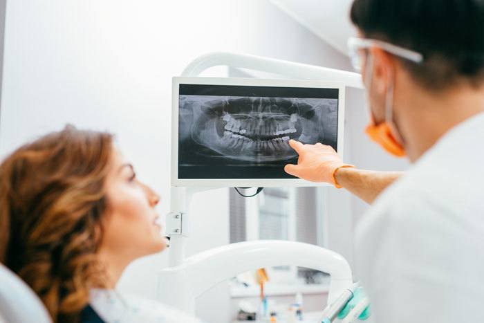
Once the x-ray exposure is complete, the digital sensor or film will be removed from the patient’s mouth. In the case of digital x-rays, the images are instantly transmitted to a computer screen. The dental professional can then review the images and determine if any additional x-rays are needed.
Step 5: Analysis and Diagnosis
The final step in the process is the analysis and diagnosis of the x-ray images. The dental professional will carefully examine the images to identify any abnormalities or signs of dental issues such as cavities, gum disease, impacted teeth, or jaw problems. They will use the x-ray images as a valuable tool to create an accurate treatment plan for the patient.
The Benefits of Digital X-ray of Teeth
Digital x-ray machine dental offers numerous benefits compared to traditional film x-rays.
- Reduced Radiation Exposure: Digital dental X-rays use significantly less radiation than traditional film X-rays, ensuring greater safety for patients, especially children and pregnant women who are more radiation-sensitive.
- Instant Image Capture and Transfer: Digital X-rays eliminate the waiting time for film development. Dentists can access images instantly on a computer screen, expediting diagnosis and treatment initiation.
- Enhanced Image Quality: High-resolution digital images enable dentists to zoom in and analyze minute details, leading to more precise diagnoses. Additionally, these images are easily stored, shared, and retrieved electronically, eliminating the need for physical film storage.
- Eco-Friendly Solution: Digital X-rays are environmentally friendly, as they are paperless and chemical-free, unlike traditional film X-rays that require chemicals for development. This eco-conscious approach reduces waste and promotes greener dental practices.
Digital dental X-rays revolutionize oral health diagnosis and treatment with reduced radiation, instant image access, high-quality images, and eco-friendliness, enabling precise diagnoses and swift treatment.
Key Takeaways: How Are Digital Dental X-rays Taken?
- Digital dental x-rays are taken using a special machine called a digital sensor.
- The sensor is placed inside the patient’s mouth and captures images of the teeth and jawbone.
- These images are then transferred to a computer for viewing and analysis.
- Compared to traditional film x-rays, digital x-rays are faster, more accurate, and emit less radiation.
- Digital x-rays also allow dentists to zoom in on specific areas and enhance the images for better diagnosis.
Frequently Asked Questions
What is the process for taking digital dental x-rays?
Taking digital X-ray of teeth involves inserting a sensor or camera into the mouth, connected to a computer. Real-time images are captured from various angles, allowing instant review, zooming, and enhanced analysis for efficient diagnosis and treatment planning.
Are digital dental x-rays safe?
Digital dental X-rays are indeed safe, offering up to 80-90% less radiation exposure than traditional film X-rays. They also eliminate chemical processing, reducing health risks. The precision reduces retakes, further lowering radiation exposure, making digital dental X-rays a safe and reliable diagnostic tool.
What are the advantages of digital dental x-rays?
Digital dental X-rays offer instant results, speeding up diagnosis and treatment planning. They provide high-quality images for accurate diagnosis, are easily manipulated, and eliminate physical storage, simplifying image access and sharing.
How do digital dental x-rays benefit patients?
Digital dental X-rays benefit patients through reduced radiation exposure, enhancing safety for those needing frequent X-rays. Additionally, they offer a more comfortable experience with flexible sensors for easier positioning and higher-quality images for accurate diagnosis and treatment planning.
Can digital dental x-rays detect all types of dental problems?
Digital dental X-rays effectively detect various dental issues, including cavities, infections, impacted teeth, and bone loss. However, they may not uncover some conditions requiring 3D cone beam computed tomography (CBCT). Additional imaging may be advised for accurate diagnosis in such cases.
Final Summary: How Digital Dental X-rays Revolutionize Dentistry
Digital X-ray of teeth have revolutionized dentistry by offering a safer and more efficient imaging method. Using digital sensors, they provide real-time, detailed images on a computer screen, reducing processing time and patient discomfort. These X-rays emit less radiation than traditional films, making them safer for all patients. Furthermore, digital images can be enhanced and compared to track changes, enabling early problem detection and effective treatment. This technology has become the new standard, significantly improving the way dental professionals diagnose and treat oral health issues.
Call or Book appointment online
:Ace Dental Care Alpharetta office: 678-562-1555 - Book Now
Ace Dental Care Norcross office: 770-806-1255 - Book Now
Disclaimer
This blog post was generated by artificial intelligence. The content of this post may not be accurate or complete, and should not be relied upon as a substitute for professional advice. If you have any questions about the content of this post, please contact us.
We are constantly working to improve the accuracy and quality of our AI-generated content. However, there may still be errors or inaccuracies. We apologize for any inconvenience this may cause.